1 out of 100 newborn is born with a heart defect. These heart diseases are of different types and present with varied symptoms. Some present in early newborn period and few of these present later in infancy or early childhood. These diseases can be life threatening as well.
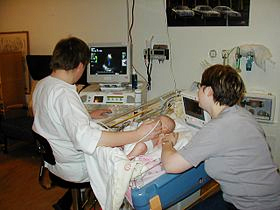
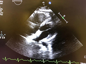
These diseases can be easily diagnosed with the help of a simple test known as echocardiography. This test is known as gold standard for diagnosis of heart defects.
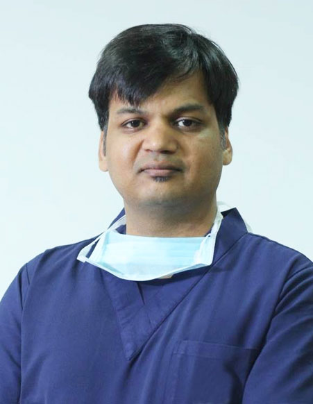
Dr Gaurav Garg has a vast experience of performing more than 25000 echocardiography on babies of every age and weight with excellent results and efficacy. He performs every kind of echocardiography on children including 2 dimensional echocardiography, 3 dimensional echocardiography and transesophageal echocardiography.
Patent ductus arteriosus (PDA) is a communication between descending Aorta and main pulmonary artery.
Ductus arteriosus is a vital structure in fetal life and it is required for survival in mother’s womb.
It closes spontaneously after few days to few weeks of birth. If it doesn’t close after birth then it is called as patent ductus arteriosus (pda).
It can cause various symptoms in the baby like
PDA can be closed by either surgery or with use of device in cath lab without surgery.
Closing of PDA in cath lab is easy and safe method of closure in experienced and skilled hands.
Device closure of PDA is a one time procedure and generally it doesn’t require re operation in whole life of baby if done successfully.
Device closure of PDA is a well known technique around the world and is preferred method of treatment over surgery.
Dr. Gaurav Garg has a vast experience of closing more than 500 PDA with device and smallest baby was 1100 grams.
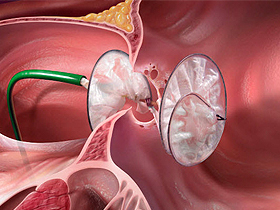
Atrial septal defect (ASD) is a common congenital heart defect and constitute around 9-10 % of all cardiac defects.
Heart has four chambers. ASD is the hole between two upper chambers of the heart. Generally, patients who are born with this hole are asymptomatic but causes symptoms in second or third decade of life.
This hole should be closed at 3-4 years of age. It can be closed by surgery or without surgery with the help of device Device closure of Asd is a very safe and effective procedure of closing Asd and is a US FDA approved procedure. It doesn’t require any cut in the body and can be safely performed through leg.
Dr Gaurav Garg has a vast experience of closing Asd with device. He has successfully done > 350 cases of Asd device closure including complicated devices also.
Ventricular septal defect (VSD) is the most common hole in the heart which lies between two lower chambers of the heart.
Babies can be symptomatic because of moderate to large VSD. Symptoms may be-
If baby is symptomatic, VSD closure should be done either surgically or with help of device in cardiac cath lab.
Device closure of VSD is a very skill full task and if operator has enough expertise, surgery can be avoided in carefully selected patients and they can get permanent cure from their disease. Patients are usually discharged home very next day of the procedure.
This procedure is not routinely performed in North India where most of the patients are sent directly for surgery in which hole is closed after cutting the chest and stopping the heart.
Dr. Gaurav Garg has successfully performed > 150 cases of VSD device closure with excellent outcomes.
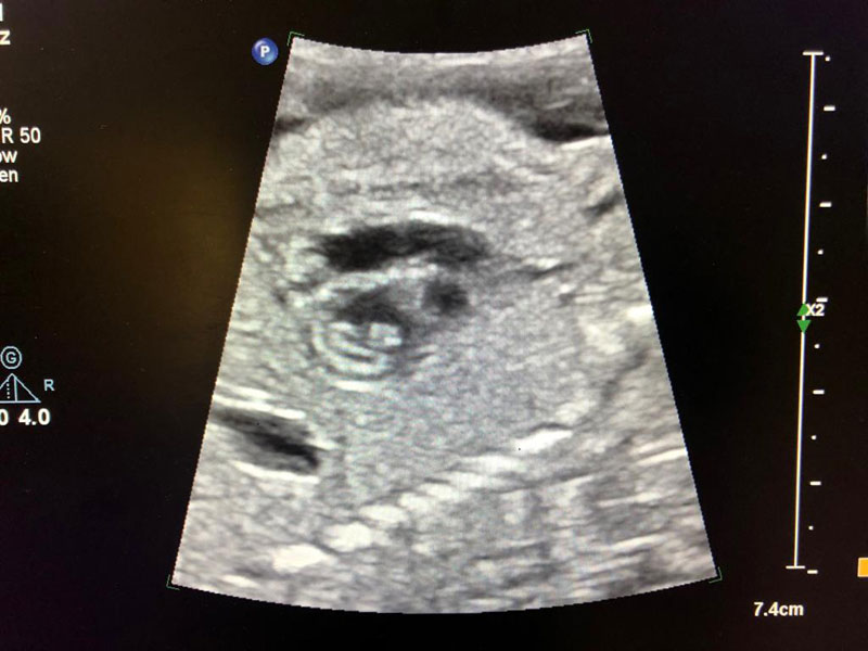
Cardiac activity of baby in mother’s womb can be seen at 22 days of gestation. By 12 weeks, heart of the baby is fully formed and functional in the mother’s womb. With the use of fetal echocardiography, we can see the structure and function of the baby’s heart. Any problem in the baby’s heart can be detected with 80-90% efficacy.
Ideal gestational age for performing this test is between 22-24 weeks of pregnancy but it can also be performed safely between 18-20 weeks Use of fetal echocardiography
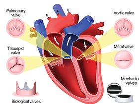
Aortic valve is the main valve which lies on left side of heart. Normally it has three leaflets which allow the blood to flow only in one direction i.e from heart to the body. When the leaflets open, blood can flow through the valve but when they close, there is no blood flow through the valve.
If the valve has 2 leaflets, the opening mechanism is affected and there can be narrowing in the valve.
This valve can be opened with the help of balloon which is passed through a peripheral vessel and is inflated across the valve.
Although this procedure is a high risk procedure but can be life saving for the baby as well.
Similarly, pulmonary valve which lies on the right side of heart can be defective and narrow.
It can cause right heart failure, breathlessness and sudden death as well.
It can also be dilated successfully in the cath lab without surgery.
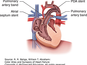
Patent ductus arteriosus (PDA) is a structure which is present in all the babies before birth. After birth, it is automatically closed within few days.
In few diseases of heart, pda is required for survival, otherwise the baby won’t survive.
This is known as pda dependent circulation.
In these newborn babies we need to keep the pda open otherwise a surgical shunt is needed which is known as BT shunt.
Pda is kept open by putting stent in the pda in cath lab without surgery.
Pda stenting is a very high risk procedure which requires a lot of skills but if it is done by experienced operator, wonderful results can be achieved and surgery can be avoided which also carries a lot of risks in these small babies.
Dr. Gaurav Garg has a vast experience of doing pda stenting in small babies.
He has published a case series of pda stenting in babies less than 2 kg body weight which is probably the first of it’s kind in India.
Copyright © 2020 Dr. Gaurav Garg All Rights Reserved | Healthcare Web Design Agency Medkeon